WHAT ARE THE COMPLICATIONS ASSOCIATED WITH DVT?
There are two serious complications associated with DVT.
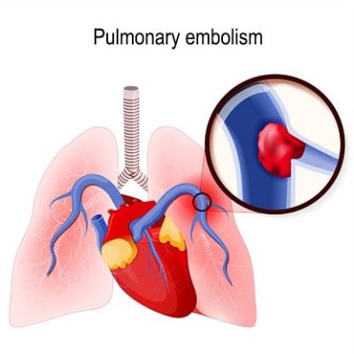
1. PULMONARY EMBOLISM (PE):
A pulmonary embolism occurs when a blood vessel in your lung becomes blocked by a blood clot (thrombus). The clot travels to your lung from another part of your body. A PE can be life-threatening! Therefore, it is essential to watch out for signs and symptoms and seek medical attention immediately if any occur.
The signs and symptoms of PE include:
Sudden shortness of breath
Chest pain or discomfort that worsens when you take a deep breath or when you cough
Fainting or feeling lightheaded or dizzy
Having a rapid pulse
Coughing up blood

2. POSTPHLEBITIC SYNDROME:
Postphlebitic Syndrome (also called Posthrombotic Syndrome) is a common complication from DVT. The damage inflicted onto the veins from the blood clot reduces blood flow in affected areas, causing:
Persistent swelling of the legs (aka edema)
Leg pain
Skin discoloration
Skin sores and ulcers
This syndrome is under-diagnosed but a common byproduct of having DVT treated with blood thinners alone. Permanent irreversible damage occurs in the affected veins and their valves because the clot remains in the impacted vein. Nearly 60-70 percent of people with DVT can develop this syndrome within two months.
WHICH DIAGNOSTIC TESTS ARE USED TO DIAGNOSIS DVT?
ULTRASOUND:
Our Ultrasound Technologist at NEEC will perform a painless and quick diagnostic test using ultrasound. A transducer from the ultrasound machine is placed over your body where there is a blood clot. As the sound waves travel through your tissue and reflect back, a computer transforms the waves into a moving image on a video screen. This visual image helps to diagnose, map, and measure the blood flow through your veins and help find any clots that might be blocking the flow. You may have a series of ultrasounds over the course of several days/weeks to determine whether a blood clot is growing or to check for a new one.

BLOOD TESTS:
Most patients with severe DVT have an elevated blood level of a substance called D dimer. Blood tests determine this.
VENOGRAPHY:
A dye is injected into a large vein in your foot or ankle. An X-Ray creates an image of the veins in your legs and feet to look for clots.
CT OR MRI SCANS:
Either can provide visual images of your veins and might show if you have a clot. (These scans may be performed for other reasons but often reveal a clot. CT or MRI Scans are performed at your local hospital.)
ARE YOU A CANDIDATE FOR DVT DIAGNOSTIC TESTING?
You are a candidate if you are experiencing many of the DVT symptoms your doctor suspects you may have DVT, or he/she has already diagnosed you with DVT.
We Can Help!
New England Endovascular Center offers expertise, state-of-the-art imaging, and resources for the diagnosis, treatment, and management of DVT. We customize each patient’s treatment plan to maximize the best possible outcome. We are committed to providing excellent, personal patient care.
Call our office today to schedule a consultation at 413-693-2852.

Our providers are nationally recognized experts in minimally invasive therapies for treating DVT.
We provide treatment for DVT for patients throughout Western, MA and Northern, CT.
To learn more about treatments available at New England Endovascular Center call 413-693-2852 to make an appointment.
